Home › Unlabelled › Myocarditis Ecg St Elevation / Myocarditis
Myocarditis Ecg St Elevation / Myocarditis
Myocarditis Ecg St Elevation / Myocarditis. St elevation myocardial infarction (stemi) is an acute coronary syndrome (acs). The st segment is the flat, isoelectric section of the ecg between the end of the s wave (the j point) and the beginning of the t wave. Today, the electrocardiogram (ecg) still has a major role in diagnosing and triage of patients presenting with chest pain. This is further complicated by the fact that acute myocarditis may cause elevated troponin levels (myocardial cells may die as a result of inflammation). The baseline is either the pr interval or the tp interval.
The combination of retrosternal chest pain and st elevation on ecg explains why clinicians often confuse acute pericarditis and stemi. The most important cause of st segment abnormality (elevation or depression) is myocardial ischaemia or infarction. Localization of myocardial infarction / ischemia using the ecg: The st in st elevation myocardial infarction is the portion of the heartbeat where there should be little or no electrical activity; The st segment represents the interval between ventricular depolarization and repolarization.
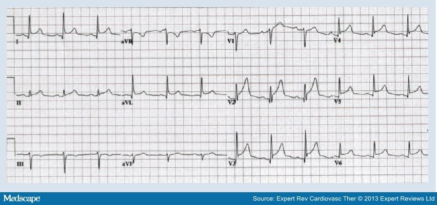
Management Of Myopericarditis from img.medscapestatic.com Both would account for a global hypokinesis on echocardiography, while st elevation in avr with diffuse st depression in the inferolateral leads is associated with left main disease. Acute myocardial infarction (mi) may be diagnosed using a 12 lead ecg. The st segment represents the interval between ventricular depolarization and repolarization. These criteria correctly classified 83% of subjects with a sensitivity of 56% and a specificity of 94%. Today, the electrocardiogram (ecg) still has a major role in diagnosing and triage of patients presenting with chest pain. The best ecg variables for the diagnosis of acute myocardial infarction were st elevation ⩾0.1 mv in ⩾1 lateral or inferior lead or st elevation ⩾0.2 mv in ⩾1 anteroseptal precordial lead. The most common ecg abnormalities seen in myocarditis are: Fulminant myocarditis is an acute, diffuse cardiac inflammation leading to cardiogenic shock, ventricular arrhythmias, and multiorgan failure.
The st segment represents the interval between ventricular depolarization and repolarization.
Both would account for a global hypokinesis on echocardiography, while st elevation in avr with diffuse st depression in the inferolateral leads is associated with left main disease. There are two types of acute coronary syndromes: It is commonly taught that such infarcts affect the basal anteroseptal myocardial segment. The st segment is the flat, isoelectric section of the ecg between the end of the s wave (the j point) and the beginning of the t wave. The combination of retrosternal chest pain and st elevation on ecg explains why clinicians often confuse acute pericarditis and stemi. The point where the end of the q wave and the st segment meet is called the j point. Localization of myocardial infarction / ischemia using the ecg: Focal myopericarditis may present with typical chest pain, st segment elevation on ekg, and biomarker elevation that may be indistinguishable from stemi. Acute myocardial infarction (mi) may be diagnosed using a 12 lead ecg. The characteristic ecg changes consistent with stemi are: Since the majority of men have st elevation of 1 mm or more in precordial leads, it is a normal finding, not a normal variant, and is designated as a male pattern; Acute myocardial infarction on ecg. Unusual cardiac risk profile, in absence of traditional cardiac risk factors, should raise suspicion of alternative diagnosis.
Patients with suspected acute st elevation myocardial infarction (stemi) are immediately referred for reperfusion therapy by either primary percutaneous coronary intervention It is commonly taught that such infarcts affect the basal anteroseptal myocardial segment. Today, the electrocardiogram (ecg) still has a major role in diagnosing and triage of patients presenting with chest pain. The best ecg variables for the diagnosis of acute myocardial infarction were st elevation ⩾0.1 mv in ⩾1 lateral or inferior lead or st elevation ⩾0.2 mv in ⩾1 anteroseptal precordial lead. Localization of myocardial infarction / ischemia using the ecg:
Acute Myocarditis Mimicking Myocardial Infarction Can Misdirect The Diagnostic Approach Sciencedirect from ars.els-cdn.com Since the majority of men have st elevation of 1 mm or more in precordial leads, it is a normal finding, not a normal variant, and is designated as a male pattern; St elevation myocardial infarction (stemi) is an acute coronary syndrome (acs). Ecg with elevation of j point in v1 and a concave upwards st elevation in v2. The st segment is the part of the ecg tracing that starts at the end of the s wave and ends at the beginning of the t wave. Focal myopericarditis may present with typical chest pain, st segment elevation on ekg, and biomarker elevation that may be indistinguishable from stemi. The baseline is either the pr interval or the tp interval. It is commonly taught that such infarcts affect the basal anteroseptal myocardial segment. The electrocardiogram is the main tool for early diagnosis for acute myocardial infarction, allowing take appropriate actions to restore, as soon as possible, the blood flow in the occluded artery.
Unusual cardiac risk profile, in absence of traditional cardiac risk factors, should raise suspicion of alternative diagnosis.
The st in st elevation myocardial infarction is the portion of the heartbeat where there should be little or no electrical activity; Both would account for a global hypokinesis on echocardiography, while st elevation in avr with diffuse st depression in the inferolateral leads is associated with left main disease. Unusual cardiac risk profile, in absence of traditional cardiac risk factors, should raise suspicion of alternative diagnosis. Acute myocardial infarction on ecg. Fulminant myocarditis is an acute, diffuse cardiac inflammation leading to cardiogenic shock, ventricular arrhythmias, and multiorgan failure. The st segment elevations in perimyocarditis are generalized, which means that they occur in the majority of the ecg leads. Our case highlights the importance of widening the physician's differential diagnostic list to include acute myocarditis in patients exhibiting pseudo. Today, the electrocardiogram (ecg) still has a major role in diagnosing and triage of patients presenting with chest pain. The st segment represents the interval between ventricular depolarization and repolarization. One of the most significant findings of myocardial infarction is the presence of st segment elevation. The point where the end of the q wave and the st segment meet is called the j point. Ecg with elevation of j point in v1 and a concave upwards st elevation in v2. There are two types of acute coronary syndromes:
It usually appears as a flat line on a ecg chart. Our case highlights the importance of widening the physician's differential diagnostic list to include acute myocarditis in patients exhibiting pseudo. The st segment is the flat, isoelectric section of the ecg between the end of the s wave (the j point) and the beginning of the t wave. Both would account for a global hypokinesis on echocardiography, while st elevation in avr with diffuse st depression in the inferolateral leads is associated with left main disease. The combination of retrosternal chest pain and st elevation on ecg explains why clinicians often confuse acute pericarditis and stemi.
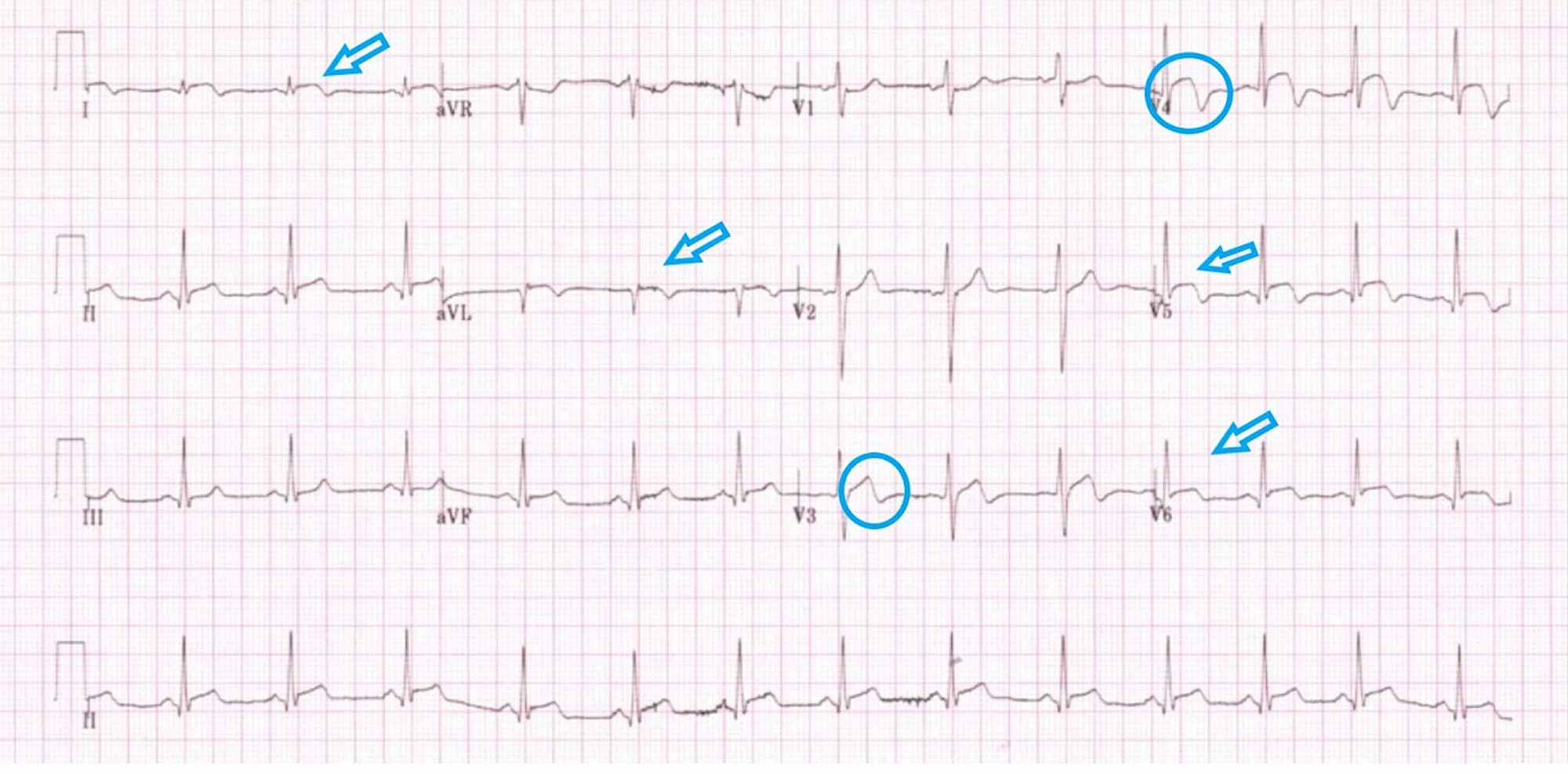
Cureus Bewildering St Elevation With Wellens Electrocardiogram Pattern Is Myocarditis In Your Differentials from assets.cureus.com The electrocardiogram is the main tool for early diagnosis for acute myocardial infarction, allowing take appropriate actions to restore, as soon as possible, the blood flow in the occluded artery. This is further complicated by the fact that acute myocarditis may cause elevated troponin levels (myocardial cells may die as a result of inflammation). As discussed previously, stemi is the result of a complete and proximal occlusion in a. Cardiac mri is useful in distinguishing myopericarditis from mi. A usually benign ecg pattern with an incidence of 5 to 13% of people so very common especially in young healthy athletes st elevation (j point elevation) of 1 mm or more in 2 or more contiguous leads (usually inferior or lateral or both) st morphology similar to pericarditis The st segment elevations in perimyocarditis are generalized, which means that they occur in the majority of the ecg leads. The best ecg variables for the diagnosis of acute myocardial infarction were st elevation ⩾0.1 mv in ⩾1 lateral or inferior lead or st elevation ⩾0.2 mv in ⩾1 anteroseptal precordial lead. The st in st elevation myocardial infarction is the portion of the heartbeat where there should be little or no electrical activity;
This is further complicated by the fact that acute myocarditis may cause elevated troponin levels (myocardial cells may die as a result of inflammation).
The characteristic ecg changes consistent with stemi are: This is further complicated by the fact that acute myocarditis may cause elevated troponin levels (myocardial cells may die as a result of inflammation). Patients with suspected acute st elevation myocardial infarction (stemi) are immediately referred for reperfusion therapy by either primary percutaneous coronary intervention Fulminant myocarditis is an acute, diffuse cardiac inflammation leading to cardiogenic shock, ventricular arrhythmias, and multiorgan failure. St elevation myocardial infarction (stemi) is an acute coronary syndrome (acs). The best ecg variables for the diagnosis of acute myocardial infarction were st elevation ⩾0.1 mv in ⩾1 lateral or inferior lead or st elevation ⩾0.2 mv in ⩾1 anteroseptal precordial lead. Other ecg changes are variable, and may include: The st segment elevations in perimyocarditis are generalized, which means that they occur in the majority of the ecg leads. These criteria correctly classified 83% of subjects with a sensitivity of 56% and a specificity of 94%. Cardiac mri is useful in distinguishing myopericarditis from mi. The point where the end of the q wave and the st segment meet is called the j point. Since the majority of men have st elevation of 1 mm or more in precordial leads, it is a normal finding, not a normal variant, and is designated as a male pattern; The most narrated ecg findings with acute myocarditis include diffuse st elevation or st depression, but wellen's type ecg has not been much associated with myocarditis in the literature.
Unusual cardiac risk profile, in absence of traditional cardiac risk factors, should raise suspicion of alternative diagnosis myocarditis ecg. St elevation myocardial infarction (stemi) is an acute coronary syndrome (acs).

comment 0 comments
more_vert